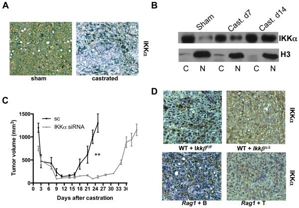
Figure 3. Role of IKKα in emergence of CR-CaP.
Tumor-bearing mice were castrated or sham operated as above. A. Tumors were analyzed one week later for nuclear IKKα by immunohistochemistry. B. Tumors removed at indicated times were divided into cytosolic (C) and nuclear (N) fractions and IKKα and histone H3 distribution was determined. C. Tumors were established using myc-CaP cells transduced with lentiviruses expressing scrambled siRNA (sc) or IKKα-specific siRNA. Mice were castrated as above and tumor volume was measured. Results are averages ± s.e.m. (n=10). P values were determined and are indicated as above. D. Tumors were established in lethally irradiated FVB males reconstituted with IkkβF/F or IkkβΔ/Δ BM or in Rag1−/− males reconstituted with either B or T cells. IkkβF/F and IkkβΔ/Δ chimeras were i.p. injected three times with poly(IC) (250 μg) prior to castration to delete IKKβ. One week after castration, tumor samples were analyzed for IKKα distribution by immunohistochemistry. Nuclear IKKα results in punctuate staining, while cytoplasmic IKKα results in diffuse staining.

