
Figure 4.
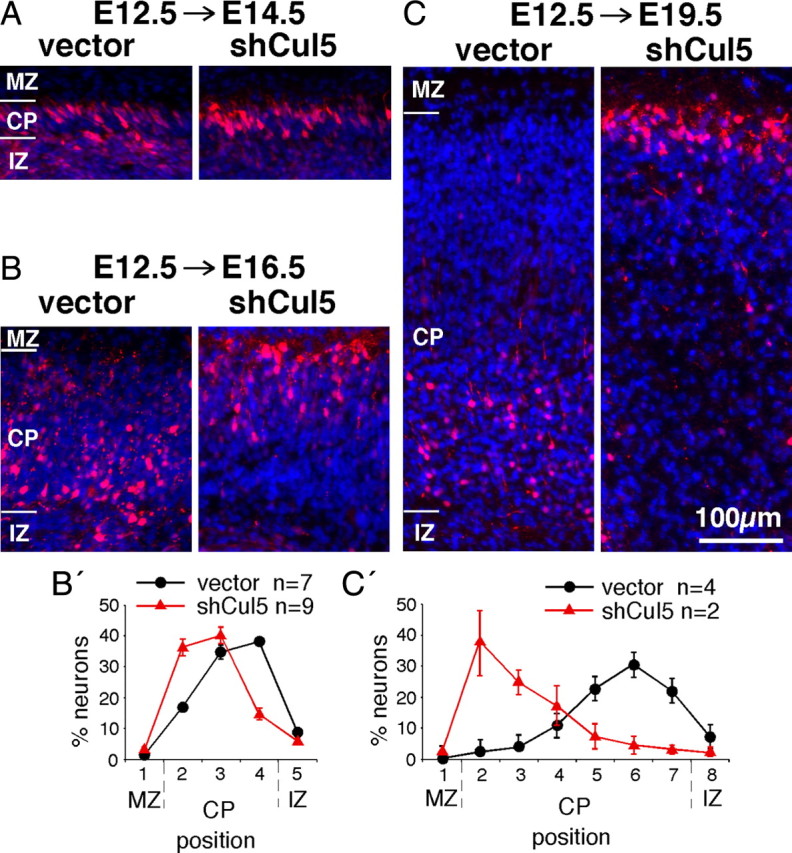
Cul5 regulates positioning of early neurons. A–C, Two micrograms of pCAG-ChFP+MFE or pCAG-ChFP-shCul5 DNA were microinjected in wild-type embryos at E12.5 and the position of ChFP-positive neurons inside the cortical plate analyzed at E14.5 (A), E16.5 (B), or E19.5 (C). Nuclei were stained with DAPI (blue). MZ indicates marginal zone; CP, cortical plate; IZ, intermediate zone. All images at same scale, bar for A–C, 100 μm. B′, C′, Neuron positions at E16.5 (B′) or E19.5 (C′). Mean ± SEM. Bin sizes are ∼50 μm. Note that control neurons remain 50–100 μm from the bottom of the CP between E16.5 and E19.5, whereas shCul5 neurons keep pace with the top of the CP as the CP expands.