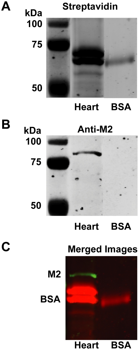
Figure 6. The organophosphorus fluorophosphonate (FP) probe does not phosphorylate M2 muscarinic receptors in guinea pig heart.
Guinea pig heart cell membrane preparations with abundant expression of M2 muscarinic receptors or BSA were reacted with a fluorophosphonate tethered to a biotin group (FP-biotin) for 24 hr prior to separation by acrylamide gel electrophoresis. Blots of these gels were probed with streptavidin tagged with an infrared fluorophore (A, red in the merged image in panel C) to localize the biotin tag and with anti-M2 receptor antibody conjugated to a different infrared fluorophore (B, green in the merged image in panel C). As indicated in the merged image (C), proteins in the heart membranes that were biotinylated by the FP probe (red) did not co-localize with bands recognized by the anti-M2 receptor antibodies (green).

