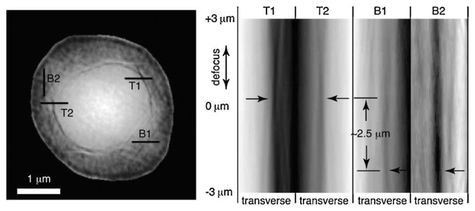
FIG. 3.
Through-focus imaging using the complex wave front reconstructed in XDM. Four selected transverse lines are shown on the magnitude-only representation of the 0 μm defocus reconstructed wave front at left. The image at right shows transverse line profiles of the reconstructed image magnitude as the wave field is defocused by propagation from the reconstruction plane. In-focus edges look like the waist of an hourglass in such a representation; the line profiles from the inner and outer spherical objects in the reconstruction appear to be at different focal planes, consistent with the interpretation that the larger parent cell is at lines B1 and B2, and the bud is at lines T1 and T2.

