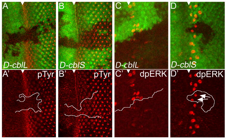
Figure 5. D-CblL negatively regulates EGFR activity.
Heat shock-induced expression of D-cblL (marked by absence of GFP) reduces pTyr labeling (in red) (A,A′) and dpERK protein (red) levels (C,C′) in 3rd instar eye discs. The top panels of each experiment are the combined GFP and pTyr/dpERK channels, the (′) panels are the pTyr/dpERK labelings only. Heat shock-induced clones expressing D-cblS do not affect pTyr (B,B′) and dpERK levels (D,D′). The white triangle marks the position of the MF. White lines in (A′,B′,C′,D′) mark the clone boundaries. The arrows in (D′) point to dpERK positive cells in D-cblS expressing clones. The UAS-D-cblL (line A18) and UAS-D-cblS (line A1) transgenes were used.

