Fig 1.
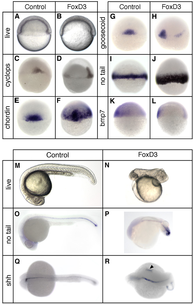
Foxd3 induction of dorsal mesoderm and axial dorsalization. At the one-cell stage embryos were injected with 25pg of foxd3 mRNA and analyzed at the shield stage (6hpf) (A–L) or at 24 hpf (M–R). (A,B) Live embryos (lateral views, dorsal right) showing the shield and blastoderm structure in uninjected (A) and foxd3-injected (B) embryos. In situ hybridization of uninjected (C,E,G,I,K) and foxd3-injected (D,F,H,J,K) embryos showing expression of cyclops (C,D), chordin (E,F), goosecoid (G,H), no tail (I,J), and bmp7 (K,L). Views shown are dorsal (C,E,F,G,I,J), dorsal lateral (D,H), or lateral with dorsal right (K,L). Uninjected (M,O,Q) and foxd3-injected (N,P,R) embryos at 24hpf. Shown are live embryos (M,N), and embryos analyzed by in situ hybridization for no tail (O,P) or sonic hedgehog (Shh) (Q,R) expression. Views are lateral with anterior left (M–P) or dorsal with anterior left (Q,R). Ectopic expression of sonic hedgehog was observed in foxd3-expressing embryos (R, arrowhead).

