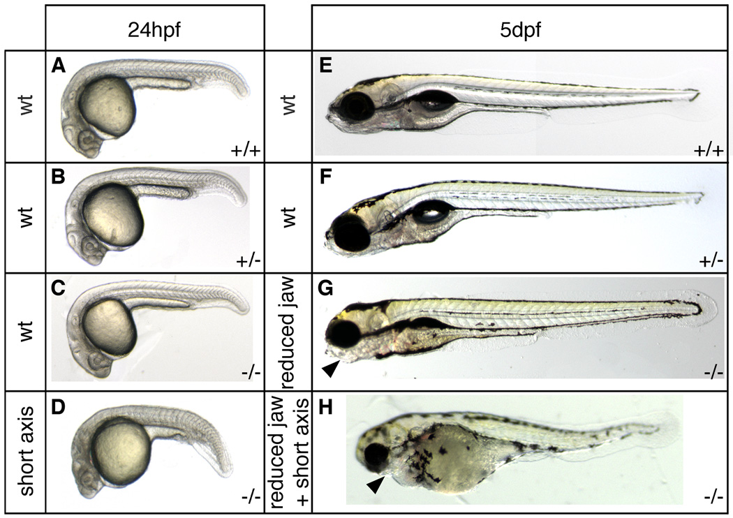
Fig. 5.
Axis formation defects in sym1 embryos. Heterozygous sym1 adults were crossed and axial development of progeny was analyzed at ~24hpf and ~5dpf. Representative samples of the two phenotypic classes observed at 24hpf (n=119): wild-type (90%) (A,B,C), and axial defects (10%) (D), and representative samples of the three phenotypic classes observed at 5dpf (n=494): wild-type (77%) (E,F), craniofacial defects (14%) (G), and craniofacial defects together with axial defects (9%) (H). Genotyping analysis indicated that at 24hpf (A–D) the wild-type phenotypic class consisted of genotypically wild-type (+/+), sym1 heterozygotes (+/−), and sym1 homozygotes (−/−), while axial defect class consisted only of sym1 homozygotes (−/−). At 5dpf (E–H) the wild-type phenotypic class consisted of both genotypically wild-type (+/+) and sym1 heterozygotes (+/−), while the craniofacial defect and the craniofacial with axial defect classes consisted only of sym1 homozygotes (−/−). Arrowhead indicates reduced jaw structures in the craniofacial defect class (G) and in the craniofacial defects together with axial defect class (H). Phenotypic class is indicated to the left of each panel and genotype is shown on the bottom right of each panel.

