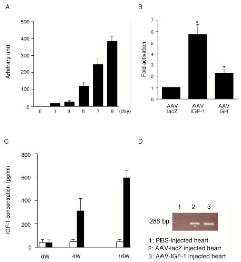
Fig. 1.
(A) IGF-1 gene expression induced with AAV in cultured cells. After infection of AAV-CMV-IGF-1 into rat H9C2 cells under the indicated time course, total RNA from cells was extracted and analysed by quantitative real time (RT)-PCR using human IGF-1 primers.
(B) Rat Akt1 gene expression. AAV-CMV-lacZ, AAV-CMV-IGF-1 or AAV-CMV-GH was infected into H9C2 cells, and 7 days after infection, each mRNA was examined by RT-PCR using rat Akt1 primers. (n=4) *P<0.05 vs. AAV-lacZ.
(C) Long term expression of AAV-CMV-IGF-1. After ligation of coronary artery, total 1×10¹¹ AAV vectors were injected into myocardium. The serum concentration of human IGF-1 in AAV-infected rats was measured by ELISA. White columns show the AAV-lacZ group and black columns show the AAV-IGF-1 group.
(D) PCR amplification of rAAV genome from infected hearts 16 weeks after infection. The primers were designed for a 286-bp fragment from ITR region to CMV promoter. Lane 1, non-rAAV-infected heart; Lane 2, AAV-lacZ infected heart; Lane 3, AAV-IGF-1 infected heart.

