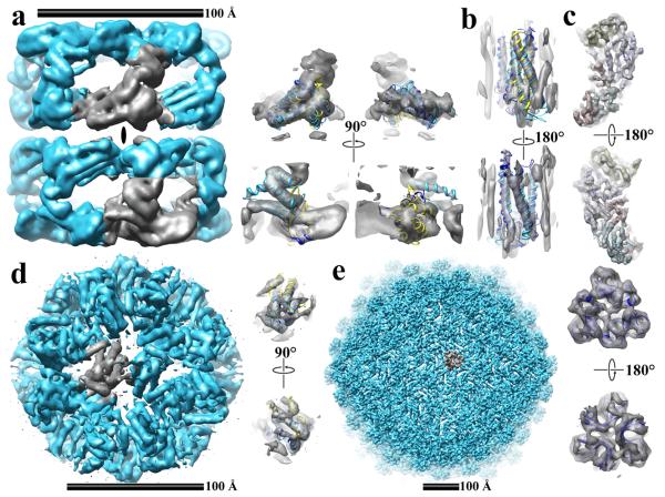
Figure 2.
Surface and ribbon representations of cryo-EM image reconstructions with known atomic structures. Shown are the image reconstructions in cyan, the segmented densities in grey, and the top three solutions identified and fitted with FREDS as blue, cyan, and yellow ribbon cartoons. A) GroEL at 6.0 Å resolution. An ellipse identifies the 2-fold symmetry axis of GroEL. B) Bovine Metarhodopsin at 6.0 Å resolution, C) the P3 subunit of Rice Dwarf Virus at 8.5 Å resolution. D) The Stressosome complex at 8.0 Å resolution. E) The bacteriophage Lambda at 7.0 Å resolutions.

