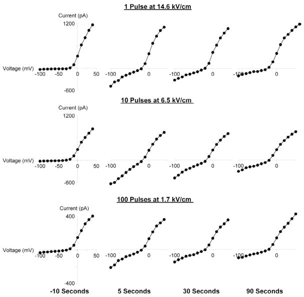
Fig. 2.
Characteristic changes in the current-voltage (I-V) function of cell plasma membrane following USEP exposure.
Shown are representative I-V curves for three cells, as recorded by a step-voltage protocol immediately prior to USEP exposure and at indicated intervals after it (See Methods for more detail). Before exposure, all cells display a subtle inward “leak” current at negative potentials and much stronger outward current at potentials between (−40) to (−30) mV due to activation of voltage-gated K+ channels. USEP exposure profoundly and reversibly enhanced the inward current component, but had little or no effect on the outward current. Note that a single pulse at a high E-field produced qualitatively the same effect as multiple pulses at lower voltages.

