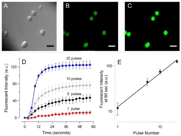
Fig. 5.
Nanosecond-duration electric pulses trigger Tl+ uptake by exposed cells.
GH3 cells were loaded with Tl+ sensitive fluorophore FluxOR™ and bathed in a solution containing 16 mM Tl+. DIC and fluorescence images were taken repeatedly every 2 sec. Exposure to a train of 1, 5, 10, or 25 60-ns pulses at 14 kV/cm started at 5 sec. A–C: representative images of cells using DIC optics (A) and fluorescence detection at 530 nm, immediately before nsEP exposure (B) and 20 sec after a train of 25 pulses (C). Calibration bar: 20 μm. (D): The amplitude and time dynamics of nsEP-induced surge in Tl+-dependent fluorescence in cells exposed to a different number of pulses (mean +/− s.e., n=3 to 5). The mean intensity of fluorescence before exposure (baseline) was subtracted from post-exposure images. (E): Same data as in D, but maximum change in Tl+-dependent fluorescence (at 60 sec post exposure) was plotted against the number of pulses in the train. Note double-logarithmic scale.

