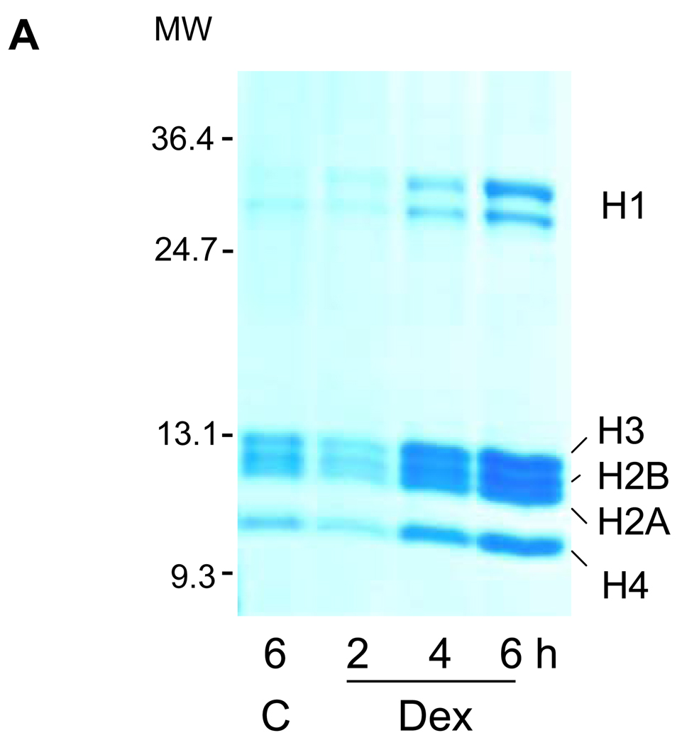
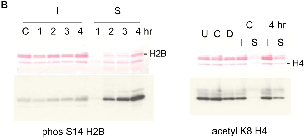
FIG. 2. Soluble chromatin contains S14 phosphorylated H2B.
(A) Histones were released during apoptotic internucleosomal degradation. Nuclei were isolated from control and dexamethasone treated cells and incubated with a lysis buffer containing 0.5% Triton X-100 at 4°C. Proteins contained in the lysis buffer were analyzed on SDS polyacrylamide gel electrophoresis and stained with 0.1% Coomassie Brilliant Blue.
(B) Thymocytes were incubated at 37°C for 1, 2, 3 and 4 hrs with 1 µM dexamethasone. Soluble (S) and insoluble (I) chromatin were isolated from nuclei. Histones were extracted from the chromatin and analyzed on SDS PAGE. left; Western blots were conducted with an anti-phosS14. C; untreated thymocytes incubated at 37°C. Proteins were stained with Ponceau-S. Right; Western blots were conducted with an acK8H4 antibody. U; untreated thymocytes on ice, C; control thymocytes, D; dexamethasone treated thymocytes at 37°C.


