
Figure 1.
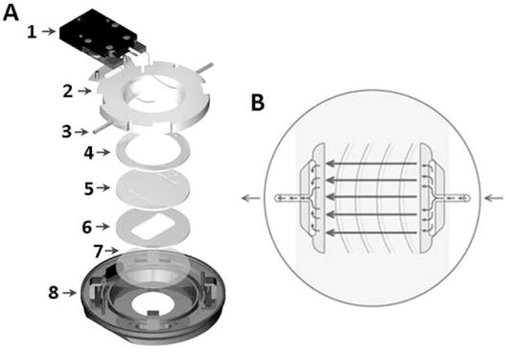
For the application of fluid flow to the cells, a closed system, parallel plate, live-cell micro-observation chamber (Focht Chamber System 2, Bioptechs Inc., Butler, PA) was utilized. (A) Exploded view of the chamber: 1-heater, 2-upper half of the chamber, 3-perfusion tubes, 4-upper gasket, 5-microaqueduct slide, 6-lower gasket, 7-coverslip, 8-lower half of the chamber which locks to the microscope stage. (B) Illustration of the microaqueduct slide perfusion technique with the laminar flow region designated by arrows.