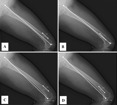
Fig. 4A–D.
The radiographic measurements of the differences are shown between (A) mechanical axis 1 versus the distal anterior cortex axis, (B) mechanical axis 1 versus the distal medullary axis, (C) mechanical axis 2 versus the distal anterior cortex axis, and (D) mechanical axis 2 versus the distal medullary axis.

