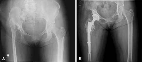
Fig. 2A–B.
(A) A preoperative anteroposterior radiograph of both hips of a 57-year-old woman shows an absent femoral head and neck with high dislocation of the greater trochanter and severe dysplastic acetabulum of the right hip and right pelvis. (B) The postoperative view obtained 10 years after surgery shows the Duraloc 1200 series cementless acetabular component is embedded well in a satisfactory position. There is a radiolucent line less than 2 mm in Zones I and III, but the acetabular component is solidly fixed. An AML femoral stem has osteolysis in Zones 1 and 7, but it is embedded solidly in a satisfactory position. The osteotomy site for subtrochanteric segmental resection is healed well.

