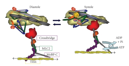
Figure 2.
Alterations in thin filament structure and promotion of the actin-cross-bridge reaction induced by Ca2+-binding to cTnC. The left panel shows the diastolic state as in Figures 1 and 2. The right panel shows the systolic state and illustrates the release of cTnI from actin and binding of the switch peptide to cTnC most likely at a hydrophobic patch induced by Ca2+ -binding to the N-lobe of cTnC. cTnT from the Tn complex on the adjacent actin strand is shown with stripes. Activation is associated with release of Tm from an immobilized state by protein signaling to cTnT and possible release of Tm from an interaction with the C-terminal end of cTnI. See text for further description.

