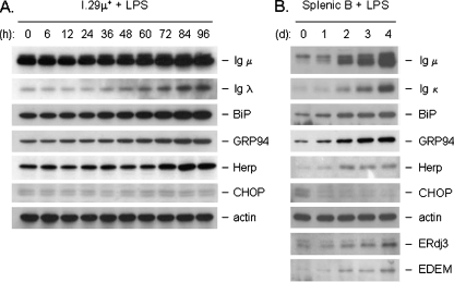
Fig. 3.
Splenic B cells show similar induction of UPR targets with LPS as I.29 μ+ cells as well as the differential induction of CHOP and Herp proteins. Cell lysates from I.29 µ+ B cells (a) or mouse splenic B cells (b) were treated with LPS for the indicated times and equal amounts of protein were loaded for each sample. After separation by SDS-PAGE and transfer, membranes were probed with the indicated antibodies. Actin served as a control for loading

