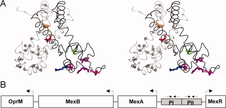
Figure 1.

Structural representation of MexR MDR mutation sites and overview of mexAB-oprM operon. A: MexR crystal structure 1LNW, represented by the chain A (black) + B (gray) dimer, which is likely to correspond to the unbound state,10 in stereo view. Mutation sites studied in this work are highlighted in chain A by displaying native sidechains in orange (L13M), red (R21W), green (G58E), blue (R70W), and magenta (T69I, R83H, R91H, L95F). Identified mutation sites in MexR leading to deficient DNA binding and/or multidrug resistance23–25,27,30,35 are indicated with Cα spheres in chain B. The figure was drawn in PyMOL.58 B: Overview of the multidrug efflux operon mexAB-oprM and mexR gene in Pseudomonas aeruginosa. The suggested MexR binding sites PI and PII in mexO are indicated.30
