
Figure 1.
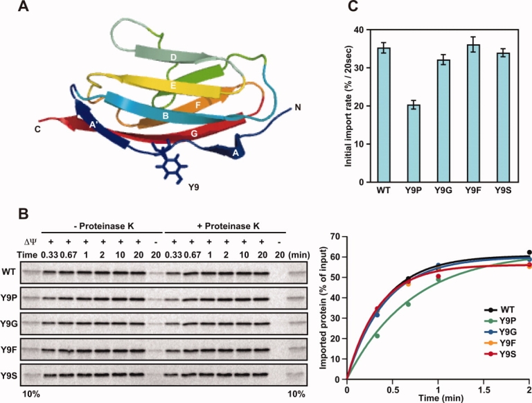
Structure of I27 and import of its Y9 mutants. (A) Cartoon diagram showing the β-sandwich structure of the I27 molecule (PDB ID, 1TIT). Tyr9 is shown in stick form. (B) Effects of mutations at residue 9 of the I27 domain on the import of radiolabeled pb2(80)-I27 fusion proteins. Mitochondria were isolated from yeast strain D273-10B. The indicated radiolabeled proteins were incubated with mitochondria at 25°C for indicated times in the absence (+ΔΨ) or presence (−ΔΨ) of valinomycin, which dissipates the membrane potential (ΔΨ) across the inner membrane. The mitochondria were treated with (+Proteinase K) or without (−Proteinase K) proteinase K, and radioactive proteins were analyzed by SDS-PAGE and radioimaging (left panels). The amounts of radiolabeled proteins added to each reaction were set to 100% and imported proteins were plotted against incubation times (right panels). 10%, ten percent of the radiolabled proteins added to each reaction. (C) Imported, proteinase-K-protected fractions in (B) were quantified, and import rates (initial slopes of the import reactions) were plotted. Values are presented as mean ± SD.