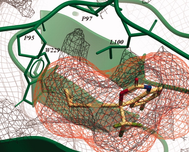
Figure 5.

The protein VdW consensus surface is shown at 10% occupancy (green wire grid) and is superimposed on the 10% occupancy NNRTI VdW consensus surface (yellow wire grid). The PDB structure 1FK9 is shown in green and with yellow carbon atoms its ligand Efavirenz. Located between the residues P95, P97, L100, and W229 is a sub-pocket unused by current NNRTIs. This sub-pocket is in fact an extension of the known cavity located between Y181, Y188, and W229.
