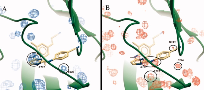
Figure 6.

Consensus surfaces visualizing all conserved HB donors surrounding the pocket (A) and all conserved HB acceptors (B) among the selection of crystal structures. For reasons of clarity, only surfaces visualizing 20% occupancy are shown. The three dimensional surfaces are superimposed on the PDB structure 2ZD1, the green ribbon indicating the backbone, and Rilpivirine depicted by yellow carbons.
