
Figure 4.
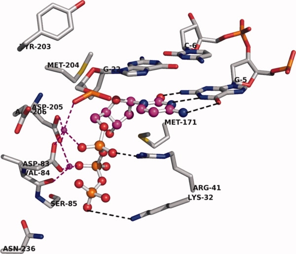
Close-up view of the active site of HDP before MD simulation. Two Mg+2 ions are shown in magenta colored small spheres. The ligand, CTP, is shown with magenta carbon in ball-and-stick mode. Mg coordination is shown with magenta dotted lines. Hydrogen bonding interactions are shown with black dotted lines. Only the important amino acids in the active site are shown; rtR41 and rtK32 are involved in ionic interactions with the phosphate group. Backbone and hydrogen atoms are removed for clarity. [Color figure can be viewed in the online issue, which is available at http://www.interscience.wiley.com.]