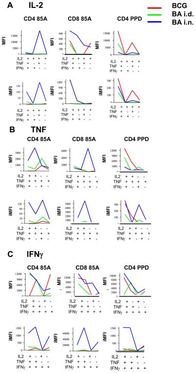
Figure 7. MFIs and iMFIs for intracellular cytokines of lung CD4 and CD8 T cells of immunized mice.
MFIs indicate the intensity of staining for intracellular cytokines in single, dual or triple cytokine producing cells. Integrated MFIs (iMFIs) are the product of the intensity of staining and the number of cytokine producing cells. MFIs and iMFIs are shown for cells producing (A) IL-2, (B) TNF and (C) IFNγ from mice immunized with BCG alone (red line) BA i.d. (green line) or BA i.n. (blue line) and restimulated in vitro with either pooled antigen 85A peptides or PPD.

