Fig. 3.

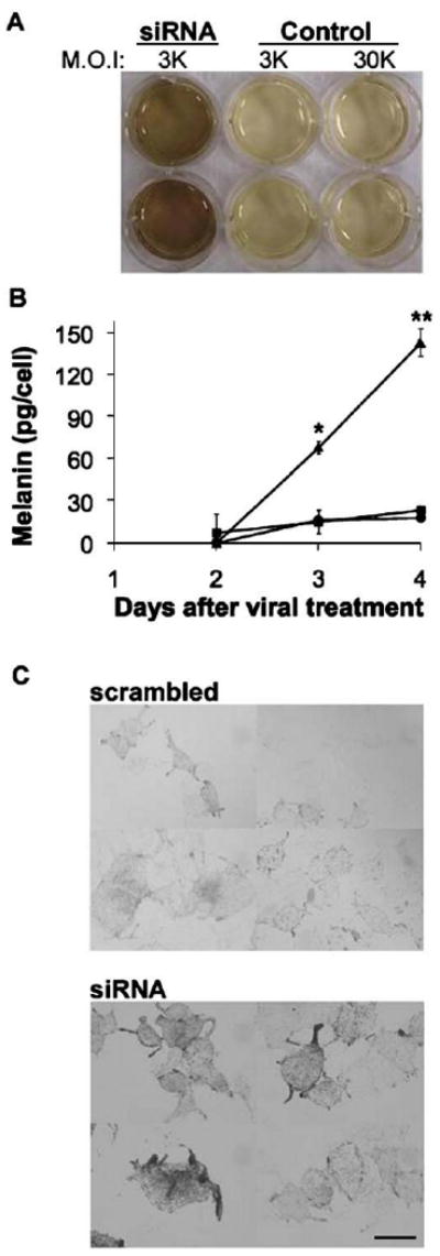
Hn1 depletion in B16.F10 cells increases melanin secretion. (A) Qualitative analysis of melanin secreted by B16.F10 cells treated with Hn1-siRNA virus at and M.O.I. of 3000 or control virus at M.O.I.s of 3000 or 30,000. The higher M.O.I. of the control virus did not impact melanin secretion. (B) Quantitative analyses of melanin secretion by B16.F10 cells treated with Hn1-siRNA (triangles), scrambled-siRNA (squares), or no virus (circles). Shown are the means of triplicate samples ±SEM (*p<0.01; **p<0.00001) of a representative experiment conducted three independent times. (C) Light microscopy of Hn1-expressing (scrambled) and -depleted (siRNA) B16.F10 cells. Scale bar: 16 μm.
