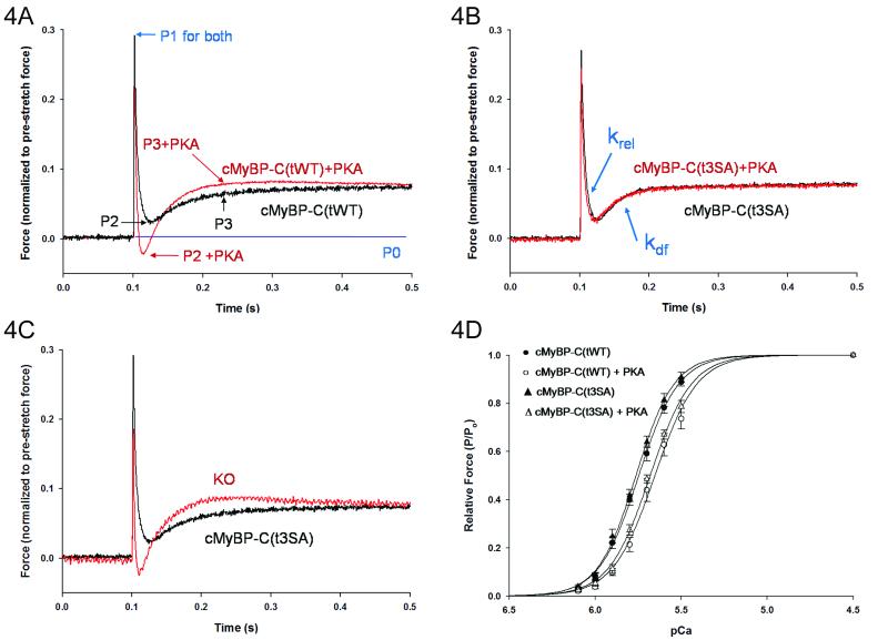
Figure 4.
PKA treatment did not affect stretch activation but decreased calcium sensitivity of force in cMyBP-C(t3SA) myocardium. (A) Forces were normalized to pre-stretch baseline P0. cMyBP-C(tWT) myocardium showed typical initial elastic response to stretch at P1, decay to P2, and delayed force development to P3 with PKA accelerating kinetics to shift P2 lower and P3 higher. (B) Stretch activation in cMyBP-C(t3SA) did not change with PKA. krel is the rate constant of force decay from P1 to P2 and kdf is the rate constant for delayed force development from P2 to P3, and (C) at baseline (no PKA treatement) cMyBP-C(t3SA) exhibited stretch activation responses similar to WT but slower than KO. (D) Normalized force-pCa plots were similar in cMyBP-C(t3SA) and cMyBP-C(tWT) myocardium at baseline and exhibited similar desensitization to calcium (right shift) after PKA treatment.

