
Figure 6.
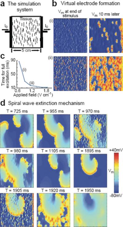
Simulation of virtual electrode formation and termination of reentry in tissue with multiple small conductivity discontinuities. a. Tissue setup schematic. Pacing stimuli are delivered to a square sheet of cardiac tissue by injecting current from the left planar electrode. Collagenous septa (short vertical lines), randomly distributed throughout the tissue, serve as conductivity discontinuities around which induced activations form in response to the applied stimuli. Septa sizes are increased 10 times and only one of every 32 is shown for clarity. Spatial resolution is 250 μm. b. Membrane potential Vm induced in the tissue by field strengths of (i) 0.8 V/cm and (ii) 1.14 V/cm (5 ms, square wave pulse). c. Time required for a single applied stimulus of various field strengths to depolarize the entire tissue from the resting state. d. Termination of reentry by eight low-voltage shocks at a cycle length corresponding to the spiral wave period. Membrane potential is shown before and after shocks #3 and 8 (S3 and S8, delivered at T= 970 ms and T= 1905 ms, respectively).