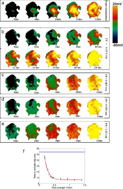
Figure 7.

Recruitment of activation sites as a function of field strength in atrial tissue. a. Propagation induced by stimulation using a point electrode. b-e. Activation of tissue by field stimulation at field strengths of 0.32, 0.46, 0.93, and 1.4 Vcm. As field strength is increased, more virtual electrodes are recruited, resulting in more rapid depolarization of the entire tissue. f. Time to full activation of the tissue as a function of electric field strength. Blue line represents time to full activation from a point stimulus.
