
Fig. 1.
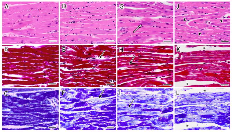
a–l Representative light photomicrographs of cardiac tissue sections stained with H&E (top panel), Masson's trichrome (center panel), and PTAH (lower panel) from female CD-1 mice necropsied at 13 week of age following transplacental exposure to vehicle (a, b, c), 3TC (d, e, f), AZT (g, h, i) or AZT/3TC (j, k, l) on days 12–18 of gestation. Heart sections from vehicle-exposed mice demonstrate characteristic features of cardiomyocytes with H&E (a) and uniform dark staining of myofibrils with trichrome and PTAH (b, c). An increase in basophilic staining (white arrow) and an attenuation of myofibers (arrowhead) was visualized with H&E staining in heart sections from NRTI-exposed mice (g,j). For trichrome and (e, h, k) and PTAH (f, i, l) staining, NRTI exposure resulted in pale or mottled (mixed staining) muscle bundles with some areas having a hyaline appearance with loss of distinct fibers and cross-striations (black arrows). Increases in endomysial space were seen most often in AZT/3TC-exposed mice (asterisks). Photomicrographs from each group represent parallel and serial sections from the same paraffin-embedded heart. Bar represents 30 μm