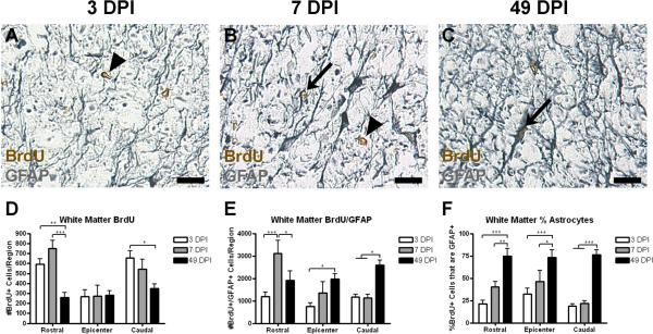
Figure 4.
In spared white matter, the number of BrdU+ cells decreases over time while the number of and percentage of BrdU+/GFAP+ cells increases. (A–C) Representative images of spared grey matter 1.0 mm caudal from the injury epicenter at 3 (A), 7 (B), and 49 DPI (C), with examples of single and double-labeled cells indicated with arrowheads and arrows, respectively. (D–F) Quantification of the number of BrdU+ cells (D), number of BrdU+/GFAP+ cells (E), and the percentage of BrdU+ cells also expressing GFAP (F) in spared white matter. Scale = 10 μm. *p < 0.05 (BrdU+ cells in the caudal region 3 vs. 49 DPI; BrdU+/GFAP+ cells in the rostral region 7 vs. 49 DPI; BrdU+/GFAP+ cells in the epicenter region 3 vs. 49 DPI; BrdU+/GFAP+ cells in the caudal region 3 and 7 vs. 49 DPI; % astrocytes in the epicenter region 7 vs. 49 DPI), **p < 0.01 (BrdU+ cells in the rostral region 3 vs. 49 DPI; % astrocytes in the rostral region 7 vs. 49 DPI), ***p < 0.001 (BrdU+ cells in the rostral region 7 vs. 49 DPI; BrdU+/GFAP+ cells in the rostral region 3 vs. 7 DPI; % astrocytes in all regions 3 vs. 49 DPI).

