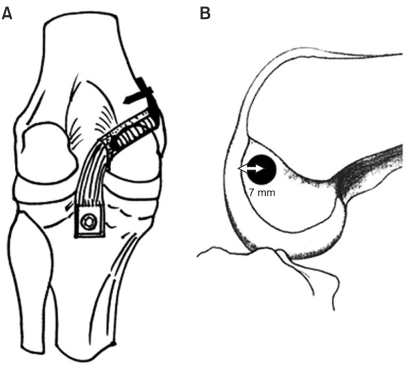
Fig. 3.
(A) The illustration shows posterior view of a single bundle tibial inlay posterior cruciate ligament reconstruction. (B) Lateral view of femoral tunnel position in the medial femoral condyle. The center of a 10 mm diameter femoral tunnel was placed 7 mm proximal to the margin of the articular cartilage of the medial femoral condyle at 11 o'clock in the left knee joint, or at 1 o'clock in the right knee joint.

