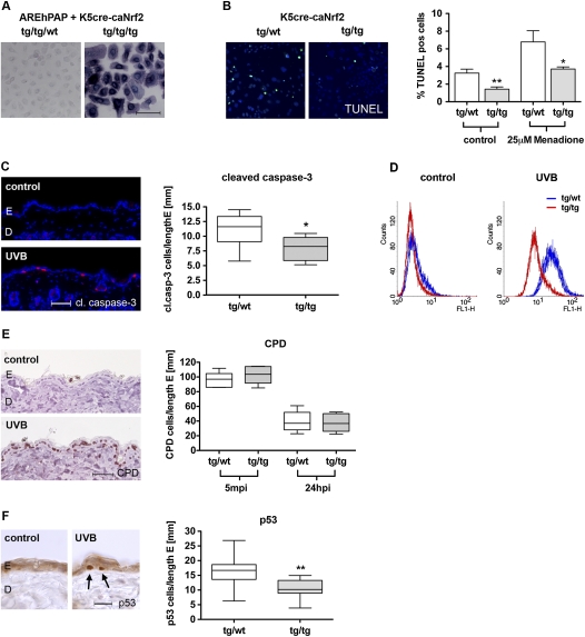
Figure 2.
ROS detoxification and protection from UVB-induced apoptosis in K5cre-caNrf2 transgenic mice. (A) hPAP activity in primary keratinocytes of double-transgenic AREhPAP/K5cre mice (tg/tg/wt) and triple-transgenic AREhPAP/K5cre-caNrf2 mice (tg/tg/tg). Bar, 100 μm. (B, left panel) TUNEL staining of primary keratinocytes (green), nuclei of tg/wild-type mice (left), and tg/tg K5cre-caNrf2 mice (right) stained with Hoechst (blue). (Right graph) Percentage of TUNEL-positive keratinocytes of tg/wild-type and tg/tg mice without (control) (P = 0.0012) and after 8 h of treatment with 25 μM menadione (P = 0.0350). (C, left) Immunofluorescence for cleaved caspase-3 (red), counterstained with Hoechst (blue), in nonirradiated (control) skin and in UVB-irradiated back skin (UVB) (24 hpi, 100 mJ/cm2) of wild-type mice. Bar, 50 μm. (Right) Number of cleaved caspase-3-positive cells per length epidermis in tg/wild-type and tg/tg mice, 24 hpi, 100 mJ/cm2 UVB (N = 9, P = 0.019). (D) FACS analysis of DCF in control (nonirradiated) and 20 mJ/cm2 UVB-irradiated immortalized keratinocytes of tg/wild-type (blue) and tg/tg (red) K5cre-caNrf2 mice. (E, left) Staining for CPD-positive cells in the epidermis of nonirradiated (control) and UVB-irradiated (UVB) wild-type mice. Bar, 50 μm. (Right) CPD-positive cells per length epidermis, 5 min (left side) and 24 hpi (right side) with 100 mJ/cm2 UVB in tg/wild-type and tg/tg K5cre-caNrf2 transgenic mice (N = 6). (F, left) Staining for p53-positive cells in the epidermis of nonirradiated (control) and UVB-irradiated (UVB) wild-type mice. Bar, 25 μm. (Right) p53-positive cells per length epidermis in tg/wild-type and tg/tg K5cre-caNrf2 transgenic mice, 24 hpi, 100 mJ/cm2 UVB (N = 11, P = 0.0039).

