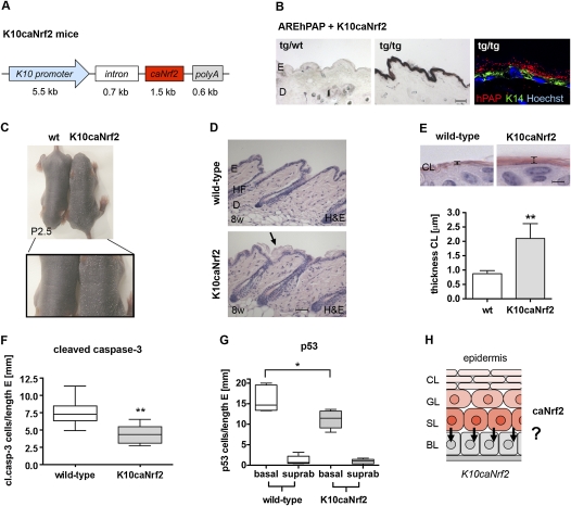
Figure 4.
Generation of K10caNrf2 transgenic mice and protection from UVB-induced apoptosis. (A) Scheme of the transgene used for generation of transgenic mice. (B) NBT/BCIP staining for hPAP activity in back skin of AREhPAP transgenic mice (left panel) and AREhPAP/K10caNrf2 double-transgenic mice (middle panel). Bar, 50 μm. (Right panel) Fluorescence staining for hPAP activity (red), immunofluorescence for K14 (green), and counterstaining with Hoechst (blue) on back skin sections of AREhPAP/K10caNrf2 mice. Note nonoverlapping localization of hPAP staining in suprabasal and K14 staining in basal keratinocytes. (C) Wild-type (wt) and K10caNrf2 transgenic mice at P2.5 in overview (top picture) and close-up (bottom picture) of lower back. Note enhanced scaling in K10caNrf2 mice. (D) Longitudinal back skin sections of wild-type (wt) and K10caNrf2 (tg) mice at 8 wk (8w). The arrow points to the area with the thicker cornified layer. Bar, 100 μm. (E, top panel) H&E staining of 2-μm sections of skin from wild-type and K10caNrf2 mice. Bar, 2 μm. (Graph) Morphometric measurement of the thickness of the cornified layer (N = 5, P = 0.0025). (F) Number of cleaved caspase-3-positive cells per length epidermis in 8- to 10-wk wild-type and K10caNrf2 mice, 24 hpi, 100 mJ/cm2 UVB (N = 8, P = 0.003). (G) Position of p53-positive keratinocytes per length epidermis in 8- to 10-wk wild-type (left) and K10caNrf2 mice (right) (N = 5, P = 0.0317). (H) Scheme of caNrf2 expression in the epidermis of K10caNrf2 mice and proposed paracrine cytoprotection of basal keratinocytes by suprabasal keratinocytes, indicated by arrows.

