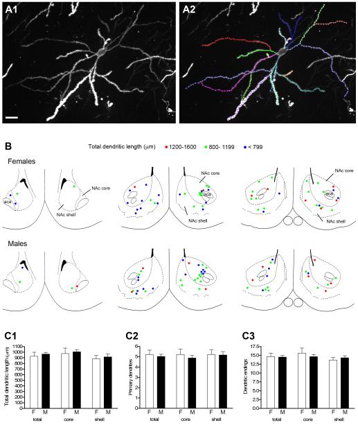
Figure 2.
A1: Projected confocal image of a DiI-labeled medium spiny neuron from the core of the NAc. A2: The same cell in A1 with individual dendritic branches traced and color coded for morphological analyses; this neuron had a total dendritic length of 1347 μm. B: Rostral to caudal (left to right) coronal sections (adapted from Paxinos and Watson, 1998) through the NAc depicting location of labeled neurons used for the whole cell analysis in males and females. Cells are represented by left and right hemisphere and color coded for total dendritic length (see key). Rostral-most section corresponds to cells imaged at approximately Bregma 2.7 mm; middle section to cells imaged from Bregma 2.2 - 1.6 mm and caudal section to cells imaged from Bregma 1.2 - 0.7 mm. C1-C3: No sex differences were found in any morphological measure including total dendritic length (C1), number of primary dendrites (C2), or number of dendritic endings (C3). Scale bar in A = 20 μm. Magenta-green version of panel 2B can be found as a supplementary figure.

