Figure 4.
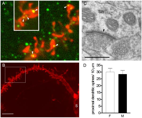
A: Single optical sections (0.2μm thickness) of a DiI-labeled dendrite (red) also immunostained for PSD-95 (green). Note that PSD-95-ir puncta were found on spine heads (arrowheads) but also on the shaft of the dendrite (inset, arrowheads). B: A lower magnification projected confocal image of the same segment of dendrite in A (indicated by white box) to illustrate the thickness of the dendritic shaft and proximity to the soma (S). C: Electron micrograph illustrating an asymmetric (excitatory) synapse (arrowhead) on a dendritic shaft (Sh) in the NAc. D: No sex difference was found in spine density on thick, proximal dendritic segments. Scale bar in B = 5 μm; in C = 0.5 μm. Magenta-green version of panel 4A can be found as a supplementary figure.

