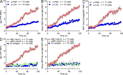
Figure 2.
Accumulation of BCRs and antigen into the contact area between B cells and antigen-containing planar lipid bilayers is affinity dependent. (A–C) The mean FI (MFI) within the contact area of Igα-YFP (A), Alexa Fluor 568–Fab anti-IgM (B), or NIP1-His12-Hylight647 (C) is given over a 120-s time course (Videos 2–4) for μ-High and μ-Low J558L cells placed on planar lipid bilayers containing NIP1-His12 (A and B) or NIP1-His12-Hylight647 (C). (D and E) MFI of either Igα-YFP (D) or Alexa Fluor 568–Fab anti-IgM (E) is given over a 120-s time course for μ-High J558L cells placed on planar lipid bilayers containing no antigen, NIP1-His12, or pNP1-His12. The data represent means ± SEM of 9–14 B cells from three independent experiments.

