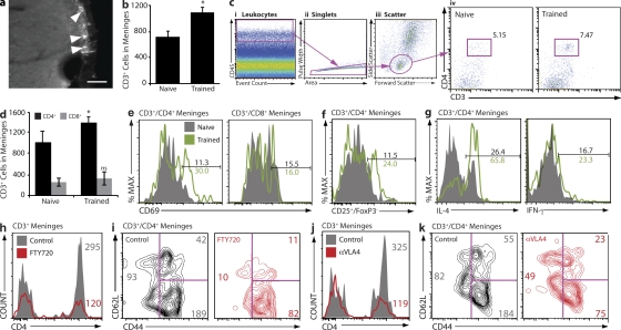
Figure 2.
Accumulation and activation of IL-4–producing T cells in the meningeal spaces of MWM-trained mice. (a and b) Whole brain cryosections were labeled for CD3. Subarachnoid spaces occupied by CD3+ cells are presented, and arrowheads point to CD3+ cells. Bar, 50 µm (a). CD3+ cells were stereologically counted. A significant increase in CD3+ cells was found in trained mice (b). (c, i–iii) Meningeal single-cell suspensions were examined by FACS for CD3+/CD4+ cells in naive and MWM-trained mice. In each sample, 104 CD45hi events were examined (i); CD45hi events were gated by scatter (ii), and CD4 versus CD3, revealing an increase in CD4+ T cells in the meninges of trained mice (iii). Experiments were performed three separate times with meninges pooled from four mice with similar results. The FACS plot shown is representative (c, iv). (d) The entirety of one single sample from each group (naive or MWM-trained) was read by FACS and normalized to one another by CD45hi count and gated on CD3, CD4, and CD8. The numbers of CD3+/CD4+ and CD3+/CD8+ cells obtained from each sample were multiplied by the total number of samples prepared from the group and divided by four (the number of mice in the group) to arrive at a representative CD4+ and CD8+ T cell count per single mouse meninges. Meningeal preparations from MWM-trained mice displayed significantly higher numbers of CD4+ but not CD8+ T cells than meningeal preparations from naive mice. Error bars represent SEM. (e and f) CD3+ cells from meninges of naive and MWM-trained mice were examined for expression of the activation markers CD69 and CD25, and the cytokines IL-4 and IFN-γ. CD4+ but not CD8+ cells from MWM-trained mice showed approximately threefold up-regulation in expression of CD69 (e) and similar up-regulation in effector (Foxp3−) T cells expressing CD25 (f) as compared with cells from naive mice. Cells from MWM-trained mice also showed a substantial increase in intracellular expression of IL-4 but not IFN-γ as compared with naive animals (g). (h–k) FACS analysis of meningeal isolates from MWM-trained FTY720-treated and control mice (h and i), and anti-VLA4–treated and control mice (j and k). Analysis of CD3+ cells from meninges of FTY720-treated mice indicates a reduction in meningeal CD4+ T cells as compared with controls; numbers indicate cell counts and reflect the analyzed sample rather than the content of the entire meningeal isolation (h); a specific reduction in CD44loCD62L+ (TNaive) subpopulation in FTY720-treated mice is presented (i). Anti-VLA4–treated mice exhibit reduction in meningeal CD4+ T cells as compared with controls (j), particularly the subpopulation characterized by the CD44hi/CD62L− (TEffector Memory) phenotype (k). At least two independent experiments with at least four mice in each group were performed. Results from independent experiments are shown. Numbers indicate percentages.

