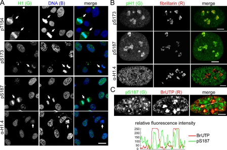
Figure 2.
Interphase phosphorylated H1.2/H1.4 are enriched in nucleoli. (A) Confocal images of asynchronous HeLa cells stained with the H1 antisera shown. DNA was stained with TO-PRO-3. Arrows indicate mitotic cells. Bar, 20 µm. (B) Confocal images of asynchronous HeLa cells costained with antisera to fibrillarin and the H1 antisera shown. Bars, 5 µm. (C) Confocal images of asynchronous HeLa cells costained with antisera to pS187-H1.4 and BrdU after pulse labeling with BrUTP to detect nascent transcripts. Bar, 5 µm. Plots of the relative fluorescence intensity of the green (pS187-H1.4) and red (BrUTP) channels along the white line shown in the merged panel demonstrate colocalization of pS187-H1.4 with BrUTP incorporation foci.

