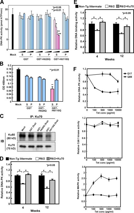
Figure 5.
Mutant Htt affects DNA–PK activity and DNA–PK complex formation in vitro and in vivo. (A) The effect of soluble/insoluble fractions of wild-type Htt (GST-Htt20Q) or mutant Htt (GST-Htt110Q) on the DNA–PK activity was examined with HeLa cell nuclear extract. Addition of the supernatant fraction (S) but not the pellet fraction (P) of mutant Htt suppressed DNA–PK activity in a dose-dependent manner (mean ± SD; n = 3). Normal Htt or GST proteins did not affect DNA–PK activity. Student’s t test confirmed the difference between GST and GST-Htt110Q. The gradient indicates that 5, 15, 50, or 150 ng of each fraction was added to 25 µl of reaction. (B) Ku70 binding to DNA was assayed as described in Materials and methods. Supernatant of GST-Htt110Q suppressed binding of Ku70 to the cleaved DNA fragments attached to the bottom of dishes. (C) IP was performed with the samples used for DNA–PK activity assay. GST-Htt110Q supernatant impaired DNA-dependent Ku70–Ku80 complex formation. (D) DNA–PK activity was assayed with nuclear extract from brain samples (cerebral cortex and striatum) of R6/2 or nontransgenic littermate (wild type) mice. Mean ± SD (n = 4) are shown. Relative activities were calculated as the ratio to the mean value of wild-type mice. Corresponding data of R6/2; Ku70 double-transgenic mice are also shown. (E) Ku70 binding to DNA was assayed with nuclear extract from brain samples of R6/2 or nontransgenic littermate (wild type) mice. Mean ± SD; n = 3. Data of R6/2; Ku70 double-transgenic mice are also shown. (F) DNA–PK, Cdk1, and MAPK/ERK activities were assayed with the Tet-on EGFP-HttQ17 and EGFP-HttQ88 stable cell lines in which induced levels of EGFP-Htt can be regulated by the concentration of tetracycline. The amount of mutant Htt necessary for impairing DNA–PK activity was titrated. Nuclear extracts from the Tet-on GFP-HttQ17 and GFP-HttQ88 stable cell lines treated with the indicated concentration of tetracyclin for 48 h were subjected to the DNA–PK assay. The induction levels of GFP-HttQ17 and GFP-HttQ88 were similar to Fig. 3 D because the two experiments were performed simultaneously. (top) An amount of mutant Htt equivalent to the endogenous Htt expression level was sufficient for inhibition of DNA–PK activity. (bottom) Meanwhile, mutant Htt did not inhibit Cdk1 and MAPK/ERK activities. These results indicated that inhibition of DNA–PK was specific. Error bars indicate SEM.

