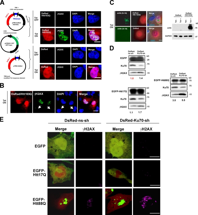
Figure 7.
Mutant Htt specifically induces γH2AX activation in a Ku70 dose-dependent manner. (A) After triple transfection of pTet-on, pTRE-DsRed-Htt103Q, and GFP-H2AX plasmids into HeLa cells, Htt expression was induced by addition of doxycycline; H2AX activation was detected by anti-γH2AX antibody. The Htt103Q/DsRed-expressing cells showed foci formation of GFP-H2AX (Siino et al., 2002) only after induction. The cells expressing only DsRed did not show foci formation even after addition of doxycycline. CMV, cytomegalovirus. Bar, 5 µm. (B) In the same field, doxycycline did not induce foci of γH2AX in nontransfected cells (arrows). Bar, 10 µm. (C) Immunocytochemistry (left) and Western blot analysis (right) show induction of Htt103Q by doxycycline. Bars, 5 µm. (D) Suppression of Ku70-enhanced activation of γH2AX. pDsRed-Ku70-shRNA and pEGFP-Htt88Q were cotransfected into HeLa cells, and immunoblots were performed with total cells. pDsRed-nonsilencing shRNA, pEGFP-Htt17Q, or pEGFP was used as control. Transfection rates of each plasmid were confirmed to be equivalent. Activation levels of H2AX (γH2AX band signals) were corrected by the band intensities of EGFP or its fusion proteins in the same blot, and the value of DsRed-nonsilencing shRNA/EGFP was defined as 1.0. (E) Confocal microscopic analysis of immunocytochemistry of γH2AX in DsRed/EGFP double-positive cells. DsRed-Ku70-shRNA–expressing cells showed higher signals of γH2AX in comparison with control cells. Bars, 5 µm.

