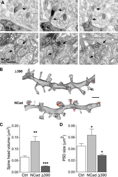
Figure 2.
Regulation of spine ultrastructure by NCad. (A) Illustrations of dendritic spines (arrows) visualized on consecutive sections obtained from a dendritic segment of a pyramidal CA1 neuron transfected with EGFP + Δ390-NCad (Δ390) or EGFP + NCad (NCad). The transfected dendritic segment is revealed by anti-EGFP immunostaining. (B) 3D reconstruction of two dendritic segments obtained from cells transfected with EGFP + Δ390-NCad or EGFP + NCad. Note the differences in size of spine heads and PSD areas (red). (C and D) Quantitative analysis of spine head volume and PSD area obtained from 3D-reconstructed dendritic segments of CA1 pyramidal neurons transfected with EGFP (Ctrl), EGFP + NCad, or EGFP + Δ390-NCad. Data are mean ± SEM of three experiments per condition (110, 58, and 127 reconstructed spines analyzed; *, P < 0.05; **, P < 0.01; ***, P < 0.001; Mann-Whitney U test). Bars: (A) 1 µm; (B) 2 µm.

