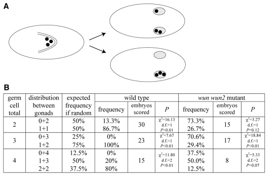
Fig. 3.
Germ cells split evenly between the embryonic gonads. (A) Cartoons representing the dorsal view of a stage 9 embryo (left) with germ cells (black circles) in the gut and stage 16 embryos (right) with germ cells in the embryonic gonads (grey ovals). In embryos containing few germ cells, the germ cells can split evenly between the two embryonic gonads (upper embryo) or all migrate into the same gonad (lower embryo), which would leave one ovary or testis devoid of germ cells in the adult. (B) Distribution of germ cells between embryonic gonads in embryos with few, but otherwise wild-type, germ cells [embryos laid by gclA26-10/Df(2R)PurP133 mothers] and germ cells lacking wunens (wun wun2 M–Z+ embryos). P value shows the probability that the observed result is significantly different from the expected distribution of germ cells if the process was random (given in the third column) using a χ2 test.

