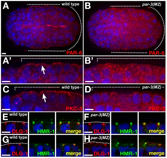
Fig. 6.
Epidermal epithelial cells in par-3(MZ) embryos. (A-B′) PAR-6 staining in the epidermis (underlying the dashed lines). The epidermis is shown at higher magnification in A′,B′. (C,D) PKC-3 staining in the epidermis. (E-H) Epidermal cells showing DLG-1 and HMR-1 in apical junctions at comma stage (E,F) or 1.5-fold stage (G,H), when junction proteins become more dispersed in par-3(MZ) than in wild-type C. elegans embryos. Scale bars: 2.5 μm.

