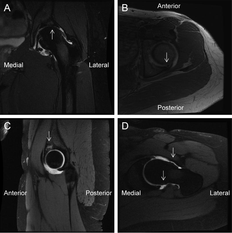
Figure 2.
Contrast magnetic resonance image of the left hip region indicating (A) the presence of severe chondrosis with broad areas of full thickness cartilage loss (T2 coronal view), (B) an associated subchondral cyst (T1 axial view), (C) a 12mm loose osteochondral body (proton dense sagittal view) and (D) large rim osteophytes on the femoral head/neck junction (T1 oblique view).

