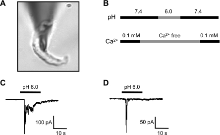
Fig. 4.
A: shape of a representative isolated cerebral VSMC. B: protocol for exposure to extracellular protons. VSMCs were briefly exposed to nominally Ca2+-free solution before exposure to extracellular H+ to prevent contraction. C and D: representative traces of the two main types of extracellular H+-evoked currents found in cerebral VSMCs.

