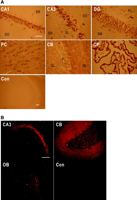
Fig. 2.
NBCn1 localization in rat brain. A: immunoperoxidase immunochemistry for NBCn1 in rat brain. Brain slices were stained with the rabbit NBCn1 antibody. Images were taken from pyramidal CA1 and CA3 regions (CA1 and CA3), dentate gyrus (DG), posterior cortex (PC), cerebellum (CB), and choroid plexus (CP). The staining without the primary antibody served as a control (Con). SR, stratum radiatum; SO, stratum oriens; SL, stratum lucidum; SM, stratum moleculare; ML, molecular layer; GL, granular layer; P, Purkinje cells. Arrows are basolateral membranes of CP epithelia. Bars represent 50 μm. The bar for CA1 applies to the other 5 panels. B: immunofluorescence immunochemistry for NBCn1 in rat brain. Brain slices were labeled with the guinea pig NBCn1 antibody and then with the Texas red-conjugated goat anti-guinea pig IgG. Images were taken from CA3, CB, and olfactory bulb (OB). Bar: 100 μm.

