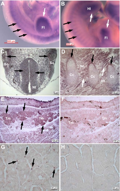
Fig. 2.
Mustn1 expression in developing and adult skeletal muscle. A and B: whole mount in situ hybridization of E10.5 and E11.5 mouse embryos, respectively, with the Mustn1 antisense probe. Black and white arrows indicate Mustn1 expression in somites and limb bud, respectively. Fl, forelimb; Hl, hindlimb. C–F: sections of an E10 (C) and E18 (D and E) day rat embryo hybridized to a Mustn1 antisense riboprobe. F: adjacent section to E but hybridized with the control sense riboprobe. Black arrows indicate Mustn1 expression in neuroepithelium and somites (So; C), perichondrium of developing ribs (D), and trapezius muscle (Tm; E). White arrows indicate Mustn1 expression in intercostal muscle (D). Nt, neural tube; Im, intercostal muscle; Cc, costal cartilage. G: adult skeletal muscle showing Mustn1 mRNA expression in nuclei at the periphery of adult myofibers, an area synonymous with the muscle satellite cell population (black arrows). H: the adjacent section to G was used as control and hybridized to the sense Mustn1 probe. Scale bar, 500 μm (A and B), 50 μm (C), 100 μm (D–F), and 20 μm (G and H).

