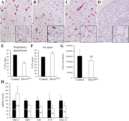
Fig. 3.
Adult Abca3Δ/Δ mice develop emphysema. Lung sections of Abca3Δ/Δ (B and D) and control littermates (A and C) were prepared from 4-wk-old (A and B) and 9-mo-old (C and D) mice and stained with hematoxylin and eosin. While no histological abnormalities were observed in 4-wk-old mice, Abca3Δ/Δ mice developed emphysema by 9 mo of age (D, inset). Photomicrographs are representative of ≥4 individual mice at each time. Scale bars, 500 μm. Magnification of inset is 3 times the original magnification. Changes in fractional areas (%Fx area) of respiratory parenchyma (E) and air space (F) were determined in 9-mo-old Abca3Δ/Δ and control littermates. Values are means ± SE. *P < 0.001. G: cell populations in bronchoalveolar lavage (BAL) fluid (BALF) from adult Abca3Δ/Δ and control littermates. BALF cell counts were similar in Abca3Δ/Δ mice and control mice. mRNAs for selected cytokines were assessed by quantitative RT-PCR in isolated BAL cells from lungs of adult Abca3Δ/Δ and control littermates and normalized to β-actin mRNA, indicating no evidence of activation of inflammation (H). Values are means ± SE of 5 animals per group. *P < 0.01 vs. control littermates. NS, no statistical difference.

