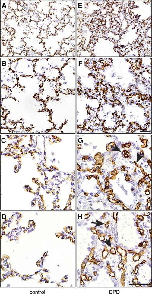
Fig. 1.
Vascular endothelial cells in the lungs of patients with bronchopulmonary dysplasia (BPD). Autopsy samples from patients that died with BPD were immunostained with antibodies against CD34 to visualize vascular endothelial cells. Age-matched controls were obtained from patients that died without clinical or pathological evidence of lung disease. A and E: low magnification. B and F: medium magnification. C, D, G, and H: high magnification. Arrows in G and H indicate dilated capillaries in BPD samples in the lung interstitium. Images are representative of 6 control and 6 BPD samples tested. Scale bar, 50 μm.

