Fig. 3.
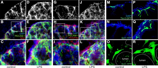
LPS stimulates vascular formation in saccular stage fetal lung explants. A–L: saccular stage fetal mouse lung explants cultured under control conditions (A–C and G–I) or in the presence of LPS (D–F and J–L) for 72 h were immunostained with antibodies against platelet endothelial cell adhesion molecule-1 (PECAM-1) and the mesenchymal markers NG2 (B, C, E, and F) or PDGF receptor-β (PDGFRβ; H, I, K, and L). Higher magnification of areas indicated by boxes in B, E, H, and K are shown in C, F, I, and L. M–R: saccular stage explants from endothelial reporter Tie2-green fluorescent protein (GFP) (M and P) or SCL-Cre-ER(T):ROSA26-enhanced yellow fluorescent protein (EYFP) (SCL-YFP; N, O, Q, and R) were cultured in control media (M–O) or in the presence of LPS (P–R). Confocal images in M, N, P, and Q show increased GFP- and YFP-positive endothelial cells in LPS-treated explants. Maximal projection of z-series of images (O and R) show increased endothelial cell sprouts and cellular processes in LPS-treated SCL-YFP explants. DAPI, 4′,6′-diamidino-2-phenylindole.

