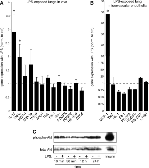
Fig. 5.
LPS stimulates inflammatory gene expression but not genes involved in growth factor-mediated vascular development. A: the effects of in vivo LPS exposure and chorioamnionitis on gene expression in fetal mouse lungs. RNA isolated from E18 mouse lungs were exposed in vivo to LPS for 72 h. Gene expression was measured by real-time PCR and compared with control RNA obtained from mice injected with saline. Only inflammatory genes were significantly increased (*P < 0.05; 6 controls and 6 LPS-exposed samples used). B: LPS increases monocyte chemoattractant protein-1 (MCP-1) expression in lung microvascular endothelial cells by real-time PCR (*P < 0.001; n = 3). No differences in endothelial markers or growth factors were detected. C: LPS did not stimulate Akt phosphorylation in lung microvascular endothelial cells. Blots shown are representative of 3 separate experiments. Insulin treatment was included as positive control. Error bars ± standard error of the mean. Ang-1, angiopoietin-1; Flk-1, fetal liver kinase-1; Flt-1, fms-like tyrosine kinase-1; HB-EGF, heparin-binding EGF-like growth factor; CTGF, connective tissue growth factor; MIP-1α, macrophage inflammatory protein-1α.

